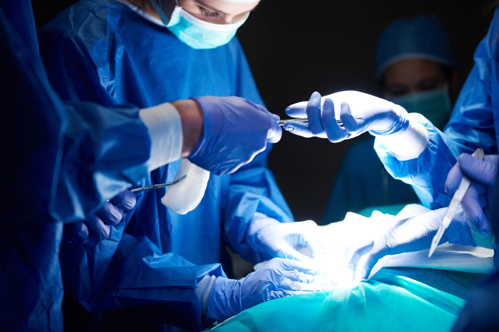
Over the last 15 years, the systematic implementation of an evidence-based perioperative care protocol (“fast-track”), has shown that hospital length of stay is reduced from 4-10 days to 1-3 days following total knee replacement(TKR) along with reduction of complications. Outpatient surgery is possible for around 15% of patients. Preoperative measures include correction of anemia, stopping smoking, intake of clear fluids till 2 hours before surgery. Standard anesthetic protocol without epidural injection with local infiltration of anesthetics around knee during surgery is advised. Medication to reduce bleeding is given before surgery. No use of tourniquet or drains or urinary catheters along with medicines to prevent post operative nausea is advised. Post operatively, patients are mobilized on same day of surgery. Once walking safely with walking aids and pain is under control are discharged usually in 1-2 days following surgery.
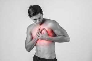
DO YOU GET CHEST PAIN & YOUR DOCTOR SAYS HEART IS NORMAL? Tietze’s syndrome is inflammation of the cartilage (Cartilage is tough but flexible connective tissue) that joins your ribs to your breastbone (sternum). This area is known as the costochondral joint. -Affects young adults under 40 – Causes infection, irritation, injury or wear and tear of cartilage -Signs and symptoms sharp chest pain made worse by 1. a particular posture, such as lying down 2.pressure on your chest, such as wearing a seatbelt or hugging someone 3. deep breathing, coughing and sneezing 4. physical activity It can be difficult to tell the difference between the chest pain associated with this condition and pain caused by more serious conditions, such as a heart attack. But a heart attack usually causes more widespread pain and additional symptoms, such as breathlessness, feeling sick and sweating. -Treatments Pain killers and occasionally steroid injection if symptoms severe -Prognosis It may improve on its own after a few weeks, although it can last for several months or more. The condition does not lead to any permanent problems, but may sometimes relapse.
ADJUSTABLE DESKS IMPROVE WORK PERFORMANCE The amount of time people spend sitting down has been linked to poor health . The study was carried out by researchers from UK and Australia. They randomised the workers in office-based clusters at University Hospitals of Leicester, who were sitting and working more than half the time to continue as usual (control group) or take part in a programme designed to reduce sitting (Stand More AT Work or SMArT programme), where adjustable desks were provided. The results showed that people in the SMArT group reduced the amount of time they spent sitting down at work. Researchers also found an increase in engagement at work, an increase in self-reported job performance & small reduction in some types of musculoskeletal problems It’s difficult to follow advice to sit down less and be more active when you work in a traditional office environment, where you are expected to work on a computer, sitting at a desk for most of the day. So the introduction of adjustable desks, which allow people to work standing up as well as sitting down, could make a big difference to a lot of people. This is an interesting study and the results show it is possible to make a fairly big difference in the time spent sitting down with the use of adjustable desks. The question now is whether this could be repeated in other workplaces https://www.bmj.com/content/363/bmj.k3870
WHAT IS INTOEING?
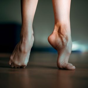
Some children’s feet turn in when they walk. This is called intoeing and is very common in young children. What problems may occur? Children who intoe may appear to trip more often at first but this soon resolves. They can be just as good at sport than anyone else. Intoeing will not affect child’s ability to walk, run or jump in the long term. What causes it? 1.Femoral anteversion: this is where the thigh bone turns inwards causing the whole leg to turn in. It is seen between 2-4 years and will resolve spontaneously by the age of 10 yrs 2.Tibial Torsion: Shin bone is twisted causing the foot to turn in. This corrects by 4-5 years as the bones grow. 3. Metatarsus Adductus: this is when the foot curves in and often results from cramped space in the womb. Most will resolve spontaneously. 4. Tight or weaker muscles The hamstrings are the muscles at the back of the thigh and tightness in these muscles can also cause intoeing. A programme of stretches helps. What can I do to help? – There is no evidence to suggest splints or special shoes will help – Encourage children not to ‘W sit’ – Out-toed activities such as ballet, horse riding, martial arts or swimming breast stroke may help Intoeing is a normal variant of development so you do not have to restrict your child’s activities.It will improve over time.
SHIN SPLINTS is the name for pain in the front of the lower legs. Common in people who do a lot of running. Symptoms The pain tends to: -begin soon after starting exercise -gradually improve when resting -dull and achy to begin with, but becomes increasingly sharp Causes Running or repetitive weight bearing which leads to inflammation of the tissue around the shin bone. Predisposing factors – sudden change in activity level – such as starting a new exercise or suddenly increasing the running distance -running on hard or uneven surfaces -poorly fitting trainers -overweight -flat feet -tight calf muscles or weak ankles Treatment -rest – stop the activity that causes pain for 2-3 weeks -ice packs for 10 minutes every few hours – painkillers -switch to low-impact activities – Eg cross-trainer or swimming and yoga You can start to return to activities over the following few weeks once the pain has gone Prevention -wear trainers with appropriate cushioning -run and train on flat, soft surfaces -introduce any changes in activity level gradually -mix high-impact exercises like running with low-impact exercises like swimming -lose weight if you’re overweight -warm up before exercising and stretch after exercising especially stretches of calves and leg muscles
CLUB FOOT is a birth defect that affects one or both feet. If baby has club foot, one or both feet points down and inwards. Club foot isn’t painful for babies, but if it isn’t treated, it can become painful and make it difficult to walk as they get older. Causes In most cases the cause of club foot isn’t known, but there may be a genetic link as it can run in families. Treatment A technique known as the Ponseti method is the main treatment for club foot nowadays. This involves gently manipulating baby’s foot into a better position, then putting it into a cast. This is repeated every week for about 5 to 8 weeks. After the last cast comes off, most babies need a minor operation to loosen the tendon at the back of their ankle (Achilles tendon). Baby then needs to wear special boots attached to each other with a bar to prevent the club foot returning. They’ll only need to wear these full-time for the first 3 months, then overnight until they’re 4 or 5 years old. Outlook Nearly all children treated with the Ponseti method will have pain-free, normal-looking feet. Most learn to walk at the usual age and enjoy physical activities, including sports, when they’re older.
DE QUERVAIN’S TENOSYNOVITIS is a painful condition affecting the tendons on the thumb side of wrist. A tendon is a strong band of tissue that attaches muscle to bone. A sheath, or covering, surrounds the tendons that go to your thumb. Tenosynovitis is an irritation of this sheath. Causes Occurs from overusing or repetitive use of thumb or wrist -playing racket sports or lifting your baby or typing in mobile Symptoms -Pain when moving thumb or wrist – Pain whilst making a fist. – Swelling and tenderness on the thumb side of your wrist -Feeling or hearing creaking as the tendon slides through its sheath. Investigations X-ray may be taken to check broken bone. Treatment -Ice pack on thumb and wrist for 20 to 30 minutes every 3 or 4 hours until the pain goes away. (Do not put ice directly next to skin as it may cause ice burn.) –Splint over wrist and thumb. -Avoid activities that worsen pain -Painkillers If things are not settling after 4-6 weeks, -Steroid injection around the tendon – Surgery for those who fail to respond to injection.
THUMB ARTHRITIS The CMC joint is at the base of the thumb, where the thumb attaches to the hand. Pain comes when the cartilage wears away. CMC OA interferes with daily activities in kitchen, around house, in people who do keyboard work, or assembly work or need to use power tools. Causes -Age -Injury -Sex: women are more likely to be affected Symptoms -Pain- At first, pain will come and go. As the arthritis advances, the pain becomes more constant and change from a dull ache to a sharp pain. -Stiffness and loss of motion -Crepitus grinding, clicking or cracking sensations -Swelling -Joint deformity -Weakness Activities that once were easy, such as opening a jar or starting the car, become difficult. Diagnosing- X ray will help Treatment Non surgical -Modify activities- Carry grocery bags over forearm instead of grasped in fingers -Hand exercises -splints – When hands are especially painful, splints can help -Heat From a warm compress help keep joints mobile. -Pain killers -Local painkiller ointments such as NSAID gels help -Steroids injected into joint provide relief for weeks to months. Surgical Treatment If nonsurgical treatment fails to give relief, surgery is done –Removal of small bone which forms the joint helps –Joint replacement
TRIGGER FINGER Trigger finger is caused by swelling of one of the tendons that run along fingers and thumbs. The swelling makes it difficult for the affected tendon to slide through its membrane (tendon sheath), causing the pain and stiffness. The swelling can cause a section of the tendon to become bunched into a small nodule at the base of the affected finger or thumb. If a nodule forms, the tendon can get trapped in the tendon sheath, causing the affected finger or thumb to become temporarily stuck in a bent position. The affected tendon may then suddenly break free, releasing finger like the release of a trigger. RISK Factors – female – 40s or 50s age – diabetes/ rheumatoid arthritis/gout – underactive thyroid SYMPTOMS – pain at the base of the affected finger or thumb – stiffness or clicking when the affected finger or thumb is moved – If the condition gets worse, finger may get stuck in a bent position and then suddenly pop straight TREATMENT In some, it will get better without treatment. However, if it isn’t treated, there’s a chance the affected finger or thumb could become permanently bent. Rest – avoiding certain activities. Pain killers Splinting – to reduce movement. Corticosteroid injections – help reduce the swelling Surgery – involves releasing the affected sheath to allow the tendon to move freely again. It’s usually used when other treatments have failed. It can be up to 100% effective.
CARPAL TUNNEL SYNDROME is pressure on a nerve in wrist. It causes tingling, numbness and pain in hand and fingers Risk factors – Overweight -do work or hobbies that repeatedly bend your wrist or grip hard -Previous injury to wrist Check if you have carpal tunnel syndrome- -pain in fingers, hand or arm -numb hands -tingling or pins and needles -a weak thumb or difficulty gripping Symptoms often start slowly and come and go. They are usually worse at night. Treatment Non surgical -wrist splint is worn on hand to keep wrist straight & it helps to relieve pressure on the nerve. Wear it at night while sleeping. – Stop or cut down on anything that causes you to frequently bend your wrist or grip hard such as using vibrating tools for work or playing an instrument. – Painkillers like paracetamol or ibuprofen may offer short-term relief -steroid injection into wrist, brings down swelling around the nerve, easing the symptoms but aren’t always a cure Surgery If symptoms are getting worse and other treatments haven’t worked, then surgery is indicated. Surgery involves an injection that numbs the wrist (local anaesthetic) and a small cut is made. The carpal tunnel inside wrist is cut so it no longer puts pressure on the nerve. The operation takes around 20 minutes and patient won’t have to stay in hospital overnight
ROTATOR CUFF TEAR Rotator cuff is a group of 4 muscles which surround the shoulder joint, which are attached to the bone via tendons. These muscles & tendons help keep the shoulder in socket & help control movements Causes The tendon/s are damaged/ torn through “wear and tear” or following an injury Symptoms -Pain at rest & at night, particularly if lying on the affected shoulder -Pain when lifting and lowering arm – Weakness when lifting arm If you have a rotator cuff tear and you keep using it despite increasing pain, you may cause further damage. Tear can get larger over time MRI/Ultrasound shows the rotator cuff tear, as well as where the tear is located within the tendon & the size of the tear Treatment Depends on age, activity level, general health & the type of tear -Rest by using a sling to help protect shoulder -Avoid activities that cause shoulder pain – Pain killers – Exercises -stretches to improve flexibility & range of motion + strengthening the muscles that support shoulder can relieve pain and prevent further injury If rest, medications & exercises do not relieve pain, an injection of a local anesthetic & steroid may be helpful Surgery is indicated if -continued pain in spite of above treatment -tear was caused by a recent, acute injury – symptoms lasting more than 6 to 12 months – significant weakness and loss of function Surgery involves re-attaching the tendon to the head of humerus (upper arm bone) by keyhole surgery or as an open procedure.
MENISCAL TEARS What are menisci? The menisci are shock absorbers that lie in the joint between the ends of the two bones in knee. In younger patients the meniscus is tough & rubbery, but can be torn in twisting injuries (traumatic tear). The meniscus becomes weaker and less elastic with age, and can be torn with minor injuries(degenerative tear) What are the symptoms? -The most common problem is pain which is felt along the joint line . Pain is worse with twisting, squatting or impact activities. -The joint may become swollen. -If the torn part of meniscus is large, locking may occur. This is caused by a large fragment getting caught in the hinge mechanism of the knee preventing the knee from straightening What is the treatment? Initial treatment is to rest the knee, ice packs and pain killer tablets. If the knee is locked and cannot be straightened, surgery is recommended to remove the torn portion caught in the knee joint. Once a meniscus is torn, it will most likely not heal on its own. In young people meniscus tear can be repaired. Repair of the meniscus is not possible in all cases. Degenerative type tears in older people are not usually repairable. Symptoms in patients with degenerative tears will settle with initial treatment. The symptoms may recur intermittently. If the symptoms continue, surgery will be required to either remove the torn portion of the meniscus or to repair the tear.
ACL Injury (Knee) The ACL is a tough band of tissue (ligament) joining the thigh bone to the shin bone. It runs through the inside of the knee and gives the knee joint stability. How common? ACL injuries are common accounting for around 40% of all sports injuries. Does all ACL injury need surgery? The decision to have knee surgery will depend on the extent of damage to your ACL and whether it’s affecting your quality of life. If the knee does not feel unstable and the patient does not have an active lifestyle, Patient may not need ACL surgery. But it’s important to be aware that delaying surgery could cause further damage to your knee. When to have the surgery? Before having ACL surgery, wait for swelling to go down and for the full range of movement to return to your knee. What happens in surgery? The ACL is reconstructed by removing what remains of the torn ligament and replacing it with a tendon from another area of the leg, such as the hamstring or patellar tendon.It is key hole procedure. Risks of surgery? ACL surgery fully restores the functioning of the knee in more than 90% of cases. In some due to other injuries to the knee, such as tears or injuries to the cartilage, knee may not be exactly like it was before the injury, and there may be some pain and swelling. Recovering from surgery usually takes around 6 months, but it could be up to a year before you’re able to return to full training for your sport.
ALL ABOUT KNEE ARTHROSCOPY What is an Arthroscopy? During an arthroscopy, a camera is inserted into the knee through two small puncture wounds. One for inserting the camera and the other for instruments. It allows us to look at the joint surfaces, cartilage and the main ligaments of the knee. It is carried out under a general anaesthetic. Once patients recover from the anaesthetic, they will be able to get up. Physiotherapist will explain exercises. Why it is done? As well as allowing to see the problem, some procedures can be performed. • Repair or removal of torn cartilages (meniscus) • Damage to the knee cap and the joint surfaces are trimmed or shaved • Small holes (micro-fractures) are made in the bone to stimulate new cartilage growth • Removal of loose bodies such as bone, cartilage or debris from the joint • If inflammation is present, samples from the lining of the joint are taken What will be the recovery following surgery? The knee may ache and swell following surgery and patient might need to take some painkillers. Majority are discharged on the same day. The majority of patients should be walking without too much pain and be able to do normal activities & office-type work within 1-2 weeks. If job involves climbing, squatting or lot of stairs, it will need two weeks to recover.Driving is possible after 5-7 days when knee. It is advisable to gradually increase the level of activity to see how knee copes.
TENNIS ELBOW is a condition that causes pain around the outside of the elbow. It usually affects adults and is more common in people who are 40-60 years of age. Men and women are equally affected. Tennis elbow is usually caused by overusing the muscles attached to your elbow and used to straighten your wrist. If the muscles and tendons are strained, tiny tears and inflammation can develop near the bony lump on the outside of your elbow. As the name suggests, tennis elbow is sometimes caused by playing tennis. However, it is often caused by other activities that place repeated stress on the elbow joint, such as decorating or playing the violin. Pain that occurs on the inner side of the elbow is often known as golfer’s elbow. You may notice pain: -on the outside of your upper forearm, just below the bend of your elbow -when lifting or bending your arm -when gripping small objects, such as a pen -when twisting your forearm, such as turning a door handle or opening a jar Treating tennis elbow It’s important that you rest your injured arm and stop doing the activity that’s causing the problem. Cold compress , painkillers and splints are used to help reduce inflammation. Some people may be offered steroid injections when other treatments haven’t worked.Steroid injections are only likely to provide short-term relief. Most cases of tennis elbow last between six months and two years. However, in about nine out of 10 cases, a full recovery is made within a year.
FROZEN SHOULDER Frozen shoulder happens when the tissue around your shoulder joint becomes inflamed.The tissue then gets tighter and shrinks which causes pain. We don’t know why this happens, though it’s more common in people with diabetes. A frozen shoulder can cause severe pain, especially at night, and you may experience sudden muscle pains, called muscle spasms The condition will usually resolve itself in time but it may take as long as two to three years. Most people with a frozen shoulder recover fully without surgery by regular stretching exercises. But if you don’t fully regain the movement in your shoulder you may need keyhole surgery to release the capsule and/or manipulation under a general anaesthetic. What are the Do’s and Dont’s? Do -follow the exercises -keep an upright posture -move your shoulder – keeping it still will make the pain worse -try heat or cold packs on your shoulder Don’t -make up your own strenuous exercises, for example gym equipment can make the pain worse -slouch when sitting – don’t roll your shoulders and bring your neck forwar
AlLL ABOUT KNEE REPLACEMENT SURGERY… Do you need knee replacement? If your knee gives pain, stiffness, instability or loss of function that affects your daily life and activities, then you will benefit from surgery
What are the advantages of Knee replacement? Freedom from pain and improved mobility and quality of life
What are the possible disadvantages? Replacement joints wearing out over time (20 years) and difficulty with some movements ( sitting cross legged or squatting)
What are the alternatives to knee replacement? We recommend non operative treatment before considering surgery including weight loss, medication,knee brace and exercises
What will be the recovery following surgery? Most people are able to start moving soon after the surgery which is good for lung function and the circulation. Physiotherapist will explain to you the exercises and help you walk. You will be discharged as soon as the wound is healing and you can safely manage to get about at home with help of walker. Most people leave the hospital between two to three days following surgery. When you are discharged, you will be given supporting therapy and follow up checks including for removal of stitches.
Dr. Gururaj S Puranik
MBBS , DNB (Trauma & Ortho)
MRCS (Edinburgh) , FRCS (Trauma & Ortho)
Fellowship in Limb reconstruction (Liverpool)
Senior Consultant – Trauma & Orthopaedics


